Retinal Diseases
Modern methods of prevention and treatment can save sight and prevent blindness from retinal disease. Screening patients for potentially blinding disorders and educating patients about symptoms of eye disease can lead to early diagnosis and a better prognosis for vision.
Retinal diseases often require urgent care. Our staff is trained to give prompt appointments to all patients with symptoms of retinal disease.
Diabetic Retinopathy, a complication of diabetes mellitus, is the leading cause of blindness among working-age Americans. Individuals with untreated diabetic retinopathy are 25 times more likely to become blind than the general population. Approximately half of all diabetic patients (approximately 16 million) in the United States do not know they have the disease.
The risk of developing diabetic retinopathy increases the longer the diabetic individual has the disease. Studies (Diabetes Control and Complication Trial) show that good blood sugar control significantly delays the onset and progression of diabetic retinopathy, as well as kidney disease, and peripheral neuropathy by greater than 50%. As visual loss usually does not occur until diabetic retinopathy is advanced, the American Diabetes Association and the American Academy of Ophthalmology recommend a thorough dilated eye examination at least yearly and more often if diabetic retinopathy is present. Laser and vitrectomy surgery can stabilize the disease and reduce the risk of visual loss.
Proliferative Diabetic Retinopathy is the formation of abnormal blood vessels on the retina. The inside of the eye behind the lens is normally filled with a clear gel-like substance called the vitreous. These abnormal blood vessels may bleed into the vitreous and obstruct vision. Laser treatment can make these dangerous blood vessels disappear, clear vitreous bleeding, and recover vision.
Non-proliferative Diabetic Retinopathy causes the normal retinal capillaries to leak. Leakage from these capillaries leads to blurring of central vision. Laser treatment of leaky capillaries significantly reduces the risk of visual loss. Other pharmacological treatments including Lucentis and Eylea are used to treat the disease.
Fluorescein Angiography and Ocular Coherence Tomography (OCT) are diagnostic tests to help the retinal specialist diagnosis and determine the extent of diseases, such as Age-Related Macular Degeneration, Diabetic Retinopathy and others. The procedures are performed in the office and are used by the retina specialist for optimal treatment.
Advanced Diabetic Retinopathy with vitreous hemorrhage (bleeding inside the eye) or retinal detachment can often be treated with vitreous surgery. Micro-instruments are introduced into the eye to remove vitreous blood and scar tissue causing retinal detachment. The surgery is usually performed in the operating room as an outpatient procedure.
Our doctors evaluate all diabetic patients at least yearly and send a written report to our patient’s family physician and endocrinologist. Our retina specialists evaluates all patients who may require treatment or close observation.
Retinal Tears are generally caused by traction of the vitreous gel on the retina. Risk factors include advancing age, myopia (nearsightedness) and previous eye surgery. Symptoms of flashing lights, floaters or a curtain (loss of side vision) suggest a retinal tear and the need for a prompt dilated eye examination. Prompt treatment of retinal tears with laser or cryotherapy (freezing treatment of the tears) can seal the tears and prevent retinal detachment.
A retinal detachment that results from a retinal tear is called a rhegmatogenous retinal detachment. The liquid vitreous that fills the eye passes through retinal tears under the retina detaching the retina from the back of the eye. If untreated, all vision will be lost.
Procedures for repairing rhegmatogenous retinal detachment include scleral buckle surgery and pneumatic retinopexy. A scleral buckle is a band placed around the eye to push the retina back to the eye wall. Pneumatic retinopexy involves the injection of a gas bubble inside the eye to push the retina back into position.
Complicated retinal detachments may require vitrectomy surgery. Micro-instruments introduced into the eye through small incisions remove scar tissue that otherwise would not allow the retina to reattach.
New instrumentation and devices including long acting perfluorocarbon gases, silicone oil, and pefluorocarbon liquids that allow the retinal specialist to reattach the retina in cases where the retina could not previously be repaired.
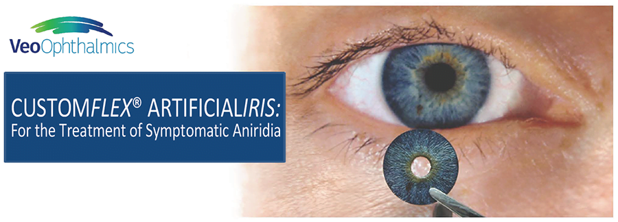
Age-related macular degeneration (AMD) is the leading cause of vision loss in Americans over the age of 50 while the cause of AMD remains unknown. There is no treatment for non-exudative or “dry” AMD but most patients with this disease retain relatively good vision. Some patients with AMD can progress to exudative or “wet” AMD where abnormal blood vessel membranes form under the macula causing bleeding and scarring resulting in severe loss of central vision. However, drugs which include Lucentis and Eylea can stabilize vision in 95% of patients with “wet” AMD and improve vision in 30-40% of cases.
Patients with AMD are asked to self monitor their vision with a checkerboard-like grid known as an Amsler grid. If decreased vision or distortion of the lines of the grid is noted, an immediate eye examination is required. Tests which include fluorescein angiography and OCT can identify these abnormal vessels and assist the retina specialist in treatment.
Many research projects are underway to determine the cause of AMD and devise new treatments. This includes vitamin and mineral supplementation (Age-Related Eye Disease Study or AREDS) as well as other drug therapies.
Macular Pucker is caused by the formation of a membrane that wrinkles the macula, the most sensitive area of the retina, thereby reducing central vision.
Macular Holes occur when the vitreous gel pulls on the macula thereby creating a hole and decreasing central vision. Both macular pucker and macular holes may be treatable with vitrectomy surgery by a retinal specialist.